
Orbital Rim
Fractures of the superior orbital wall are termed orbital rim fractures and can occur in isolation or in combination with frontal sinus fractures. Orbital rim fractures occur most commonly in isolation compared to other orbital fractures and result from direct impact on the frontal brow / supraorbital ridge.
Scrollable Stack Images
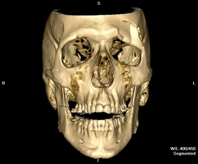
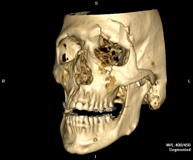
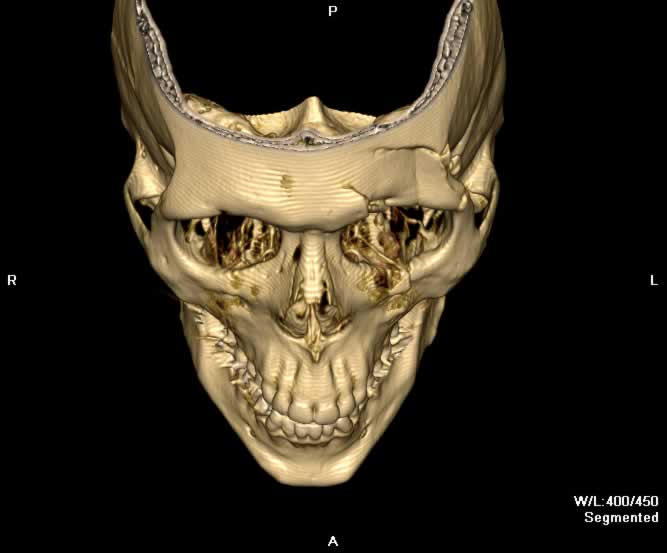
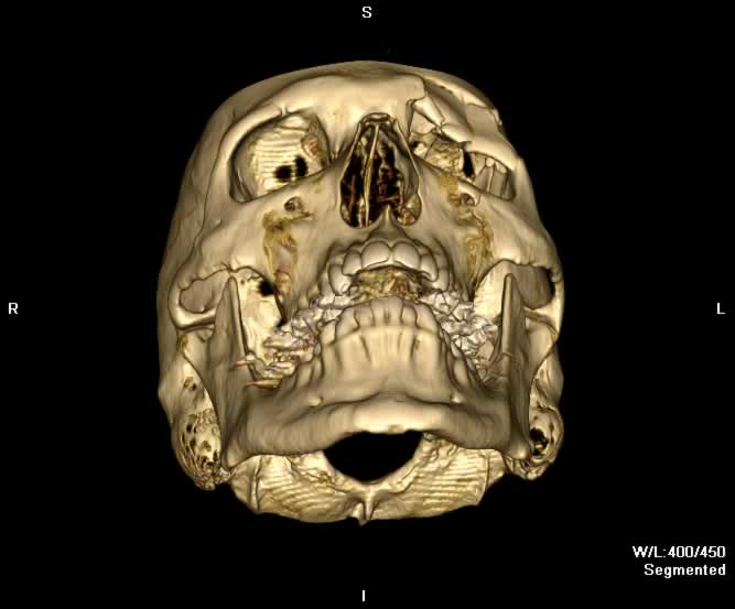
Images show an orbital rim fracture with communition and depressed of the left frontal bone. Fracture fragments of the left frontal bone are posteriorly and inferiorly displaced, extending into the intracraninal vault and the orbit. A small bony fragment impinging on the left superior rectus muscle can be appreciated. There are also fractures of the remaining walls of the left orbit - lateral, medial, and inferior. The orbital floor fracture is just lateral to the infraorbital nerve. A mild left tripod fracture, outer and inner table left frontal sinus fracutres, and displaced cribiform plate fracture are seen. The left globe and optic nerve appear intact. Hemorrhagic fluid is seen in both the left maxillary and left frontal sinuses. The pterygoid plates and mandible are intact.
Static 2D
 |
 |
 |
 |
| Click to enlarge | |||
Static 3D
 |
 |
 |
 |
| Click to enlarge | |||
Rotating 3D
Return to top
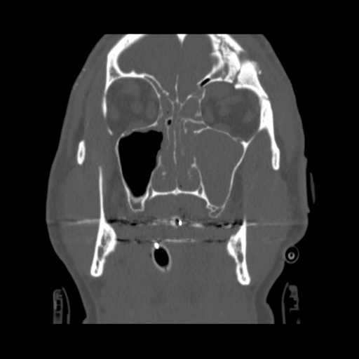
Coronal image demonstrates communition of the frontal bone with inferior displacement into the left orbit. Widening of the frontozygomatic suture is also appreciated, part of a mild left tripod fracture.
Return to top
A more posterior coronal image demonstrates a fragment from the comminuted left frontal bone impinging upon the superior rectus muscle.
Return to top
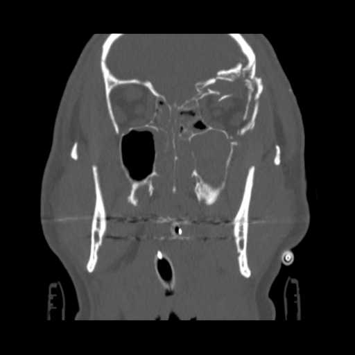
Axial image demonstrates bony fragments from the lateral orbital wall displaced into the orbit. A fracture of the medial wall of the left orbit can also be appreciated.
Return to top
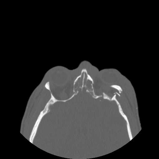
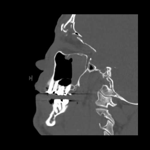
Sagittal image demonstrates fragmentation of the outer and inner tables of the left frontal bone. An inferiorly displaced bony fragment impinging on the superior rectus muscle can be faintly seen. A fracture of the cribiform plate is also visible.
Return to top
Return to top
Return to top
Return to top
Friends
 |
Frontal Sinus |
 |
Lamina Papyrecea |
 |
Orbital Floor |
 |
Orbital Blowout |
Groups
 |
Orbital Fractures |
 |
Nasal Fractures |
 |
Tripod Fractures |
 |
LeFort Fractures |
 |
Smash Fractures |
 |
Mandibular Fractures |