Orbital Floor
Orbital floor fractures can occur in isolation, or in continuation of a tripod, LeFort II, or nasoorbitoethmoid complex (NOE) fracture. Orbital floor fractures commonly result in orbital emphysema and hemorrhage into the ipsilateral maxillary sinus. Inferior rectus involvement should be assessed and may only present with subtle imaging findings in the pediatric "trapdoor" fracture pattern.
Scrollable Stack Images

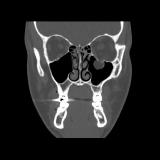
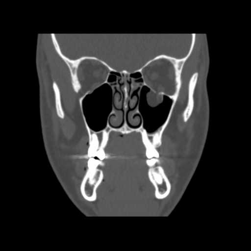
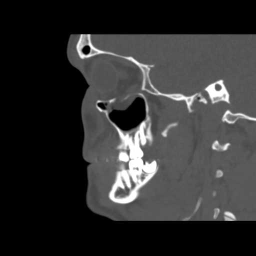
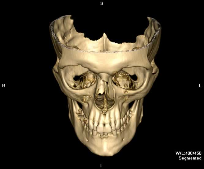
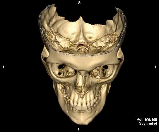
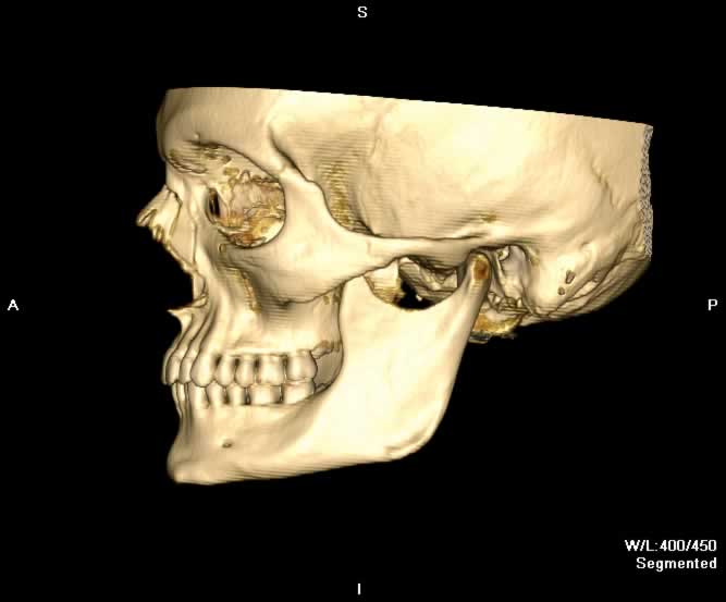
Images show a left orbital floor fracture where a portion of the inferior rectus muscle appears interposed between bony fragments. Intraconal fat herniates inferiorly through the bony defect into the left maxillary sinus. Additionally, paired nasal bone fractures can be appreciated.
Static 2D
 |
 |
 |
 |
| Click to enlarge | |||
Static 3D
 |
 |
 |
 |
| Click to enlarge | |||
Rotating 3D

Return to top
Conoral image demonstrates intraorbital fat herniating inferiorly into the left maxillary sinus through a bony defect in the orbital floor. The left inferior rectus muscle abuts the fracture site.

Return to top
An adjacent coronal image demonstrates a bony fragment from the orbit floor appearing interposed with the left inferior rectus muscle.

Return to top
Sagittal image demonstrates discontinuity of the orbital floor with herniation of the intraorbital fat into the maxillary sinus.

Return to top
Axial image demonstrates fragmentation of the left orbital floor with herniation of intraorbital fat into the maxillary sinus. Displaced paired nasal bone fractures with a small locule of subcutaneous air can also be seen.

Return to top

Return to top

Return to top

Return to top
Friends
 |
Orbital Rim |
 |
Frontal Sinus |
 |
Lamina Papyrecea |
 |
Orbital Blowout |
Groups
 |
Orbital Fractures |
 |
Nasal Fractures |
 |
Tripod Fractures |
 |
LeFort Fractures |
 |
Smash Fractures |
 |
Mandibular Fractures |