LeFort Fractures
The LeFort group is a set of three fracture patterns involving the midface. All of these fractures patterns involve the pterygoid plates. The converse is true: a fractured pterygoid plate is almost always indicative of a LeFort fracture. LeFort fractures most commonly result from low midline moderate blunt force trauma. These fracture patterns were described by Rene Le Fort, a French surgeon, through his infamous experimentation on cadavers in 1901. The three LeFort patterns are seen when blunt force trauma preferentially disrupts the horizontal rather than the vertical buttresses of the facial skeleton. Although classically described as symmetric, they frequently are not, and can be compound in nature. They are less common than they once were, since high-speed motor vehicle collisions frequently involve such great forces that they instead result in a smash pattern. Additionally, the LeFort classification does not account for two other commonly encountered fracture patterns: isolated alveolar ridge fractures and fractures of the anterior maxillary wall from focal trauma.
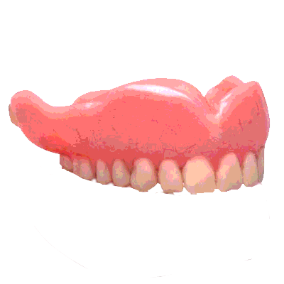
LeFort I fractures occur in the horizontal plane above the alveolar process extending from the nasal septum to the pterygoid plates, resulting in “floating palate.” On imaging, this fracture pattern can be recognized by its involvement of the anterolateral margin of the nasal fossa. On physical exam, the maxillary teeth are mobile relative to the remainder of the face.
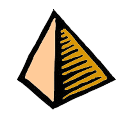
LeFort II fractures result in a pyramidal fracture extending from the maxillia to the nasal bridge, through the orbits to the pterygoid plates. The teeth are the base of the pyramid and the nasofrontal suture is the apex. This fracture pattern can be recognized by its involvement of the inferior orbital rim on imaging. A LeFort II fracture can result in the classically described “dish-face” deformity on physical exam due to posterior displacement of the free-floating maxillary teeth and nose. Infraorbital nerve injury and CSF rhinorrhea can also be seen.
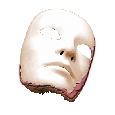
LeFort III fractures are transversely (coronally) oriented fractures extending superiorly from the maxilla to the zygomatic arches and posteriorly through the orbits to the pterygoid plates. This fracture pattern can be recognized by its involvement of the zygomatic arch on imaging. LeFort III fractures result in craniofacial dissociation manifested by mobility of the maxillary teeth, nose, and zygoma relate to the remainder of the skull. CSF rhinorrhea may also occur.
 |
LeFort I |
 |
LeFort II |
 |
LeFort III |
Friends
 |
LeFort I |
 |
LeFort II |
 |
LeFort III |
Groups
 |
Orbital Fractures |
 |
Nasal Fractures |
 |
Tripod Fractures |
 |
LeFort Fractures |
 |
Smash Fractures |
 |
Mandibular Fractures |