LeFort I
LeFort I fractures occur in the horizontal plane above the alveolar process extending from the nasal septum to the pterygoid plates, resulting in "floating palate." On imaging, this fracture pattern can be recognized by its involvement of the anterolateral margin of the nasal fossa. On physical exam, the maxillary teeth are mobile relative to the remainder of the face.
Scrollable Stack Images

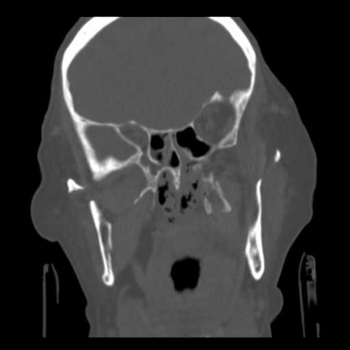
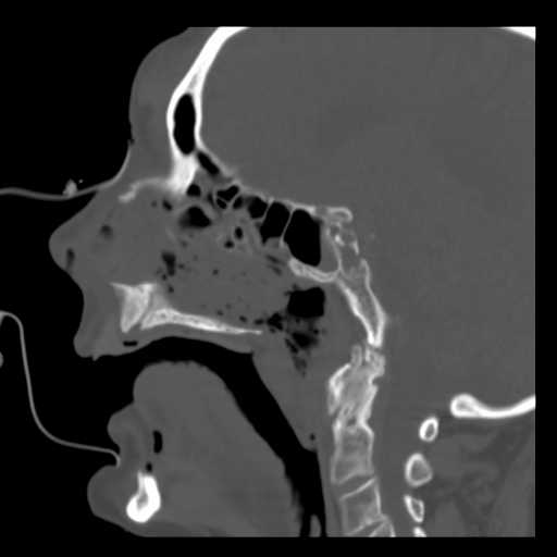
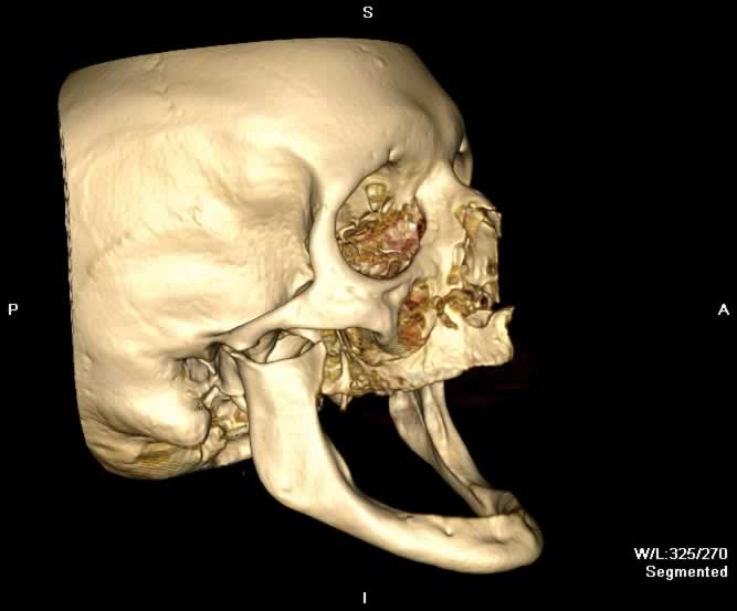
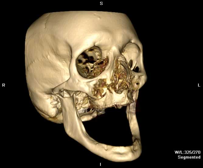
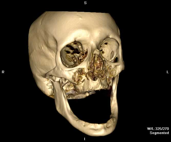
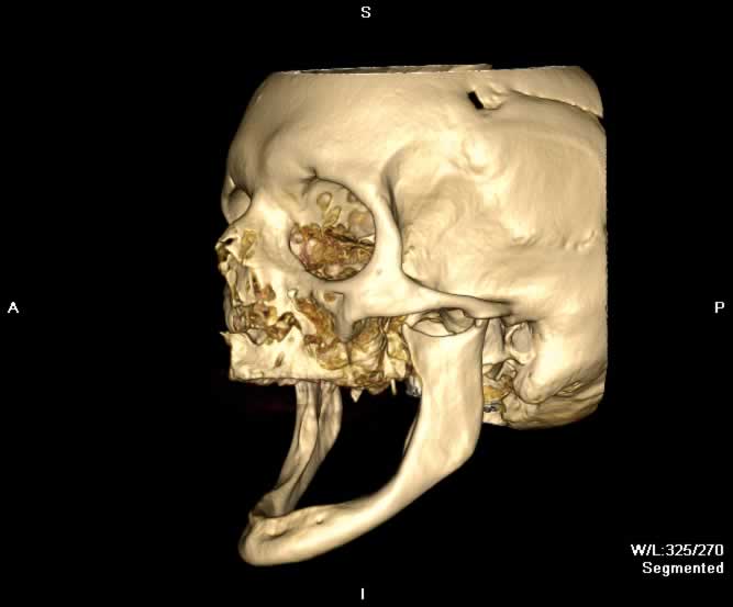
Images show bilateral LeFort I fractures. Minimally displaced fractures can be seen through the bilateral pterygoid plates and medial walls of the maxillary sinuses. Fracture through the bony attachments of the palate can be appreaciated in multiple planes. Minimally depressed nasal bone fractures are also seen.
Static 2D
 |
 |
 |
 |
| Click to enlarge | |||
Static 3D
 |
 |
 |
 |
| Click to enlarge | |||
Rotating 3D

Return to top
Axial image demonstrates fractures through the bilateral pterygoid plates.

Return to top
Coronal image demonstrates minimally displaced fractures through the anterior bony attachments of the palate - both lateral and medial walls of the inferior maxillary sinus. Hemorrhagic opacification of the ethmoid and maxillary sinuses is seen.

Return to top
A more posterior coronal image demonstrates fractures through the medial and lateral pterygoid plates bilaterally.

Return to top
Sagittal image demonstrates a midline fracture through the maxillia and the hard palate. A minimally depressed nasal bone fracture can also be appreciated.

Return to top

Return to top

Return to top

Return to top
Friends
 |
LeFort II |
 |
LeFort III |
Groups
 |
Orbital Fractures |
 |
Nasal Fractures |
 |
Tripod Fractures |
 |
LeFort Fractures |
 |
Smash Fractures |
 |
Mandibular Fractures |