
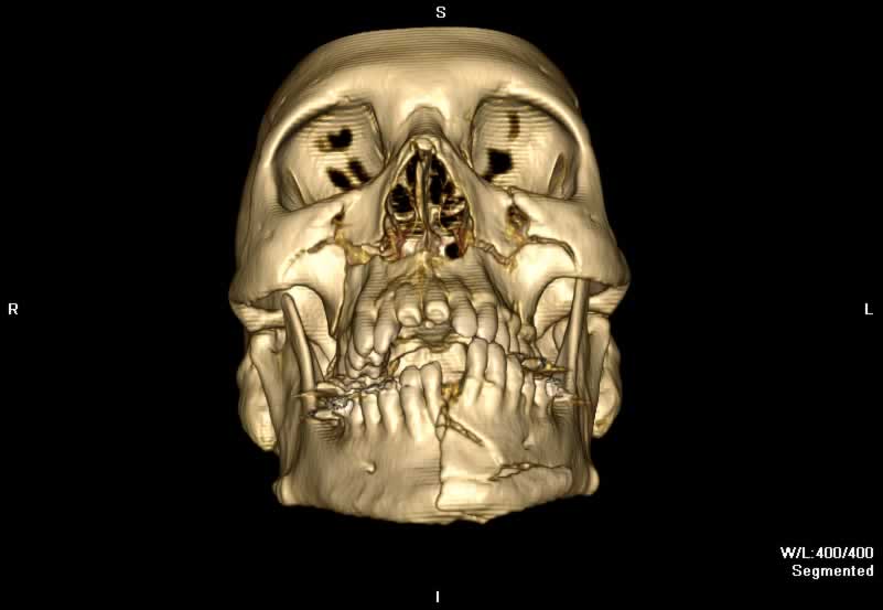
LeFort II
LeFort II fractures result in a pyramidal fracture extending from the maxillia to the nasal bridge, through the orbits to the pterygoid plates. The teeth are the base of the pyramid and the nasofrontal suture is the apex. This fracture pattern can be recognized by its involvement of the inferior orbital rim on imaging. A LeFort II fracture can result in the classically described "dish-face" deformity on physical exam due to posterior displacement of the free-floating maxillary teeth and nose. Infraorbital nerve injury and CSF rhinorrhea can also be seen.
Scrollable Stack Images
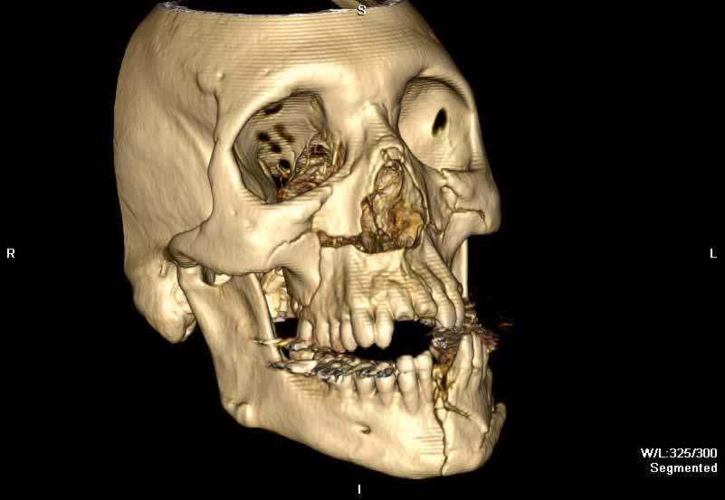
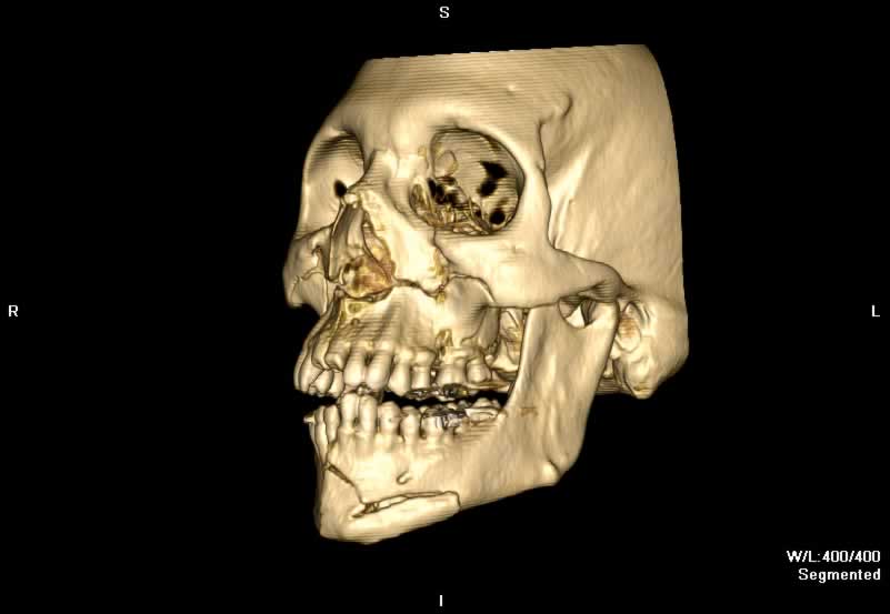
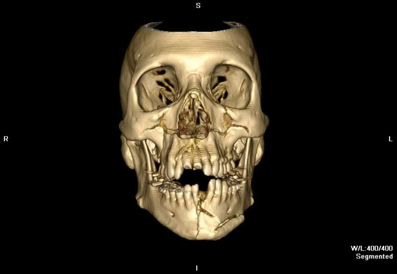
Images show bilateral LeFort II fractures. Bilateral displaced fractures of the medial and lateral pteygoid plates are seen. Fracture lines extend superiorly from the inferolateral aspects of the maxilla through the anteromedial walls of the maxillary sinuses, in close proxmity of the infraorbital foramina. Fractures of all three walls of both maxillary sinuses are seen. Fracutres of the orbital floors can be appreciated. Comminuted nasal bone and septum fractures are seen. A comminuted and displaced mandibular symphysis fracture is also seen.
Static 2D
 |
 |
 |
 |
| Click to enlarge | |||
Static 3D
 |
 |
 |
 |
| Click to enlarge | |||
Rotating 3D
Return to top
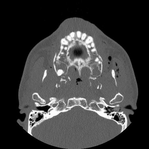
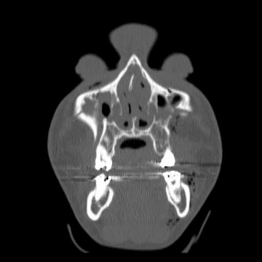
Axial image demonstrates fragmentation of the superior portions of the medial and lateral pterygoid plates bilaterally. The inferior aspect of the pyramidal fractures of the maxilla can be appreciated.
Return to top
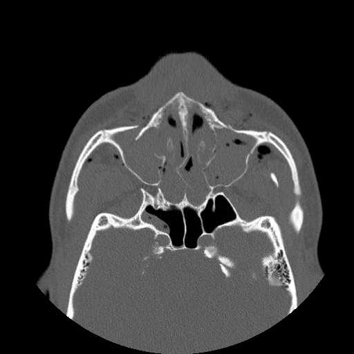
A more caphalad axial image demonstrates fractures of the anterior and medial walls of both maxillary sinuses. A fracture of the posterolateral wall of the left maxillary sinus can be appreciated. A buckle fracture of the nasal septum is seen. Hemorrhagic opacification of the ethmoid and maxillary sinuses is seen.
Return to top
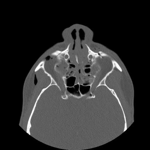
A more cephalad axial image demonstrates fractures running superomedially along the anterior maxillary walls to the nasal ridge.
Return to top
Coronal image demonstrates superomedial fractures of the maxillary walls and minimally displaced orbital floor fractures.
Return to top
Return to top
Return to top
Return to top
Friends
 |
LeFort I |
 |
LeFort III |
Groups
 |
Orbital Fractures |
 |
Nasal Fractures |
 |
Tripod Fractures |
 |
LeFort Fractures |
 |
Smash Fractures |
 |
Mandibular Fractures |