
Nasoorbitoethmoid Complex
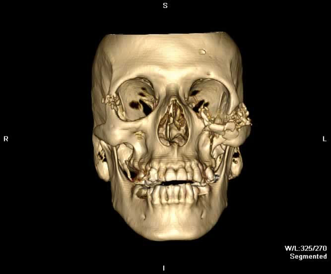
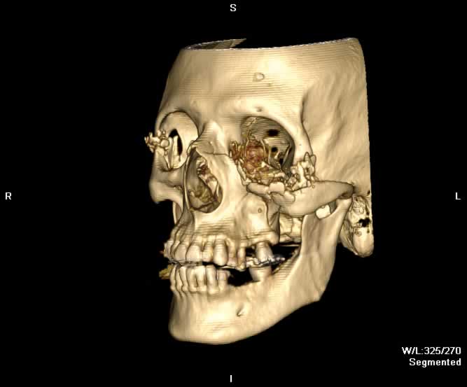
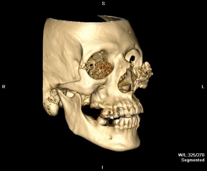
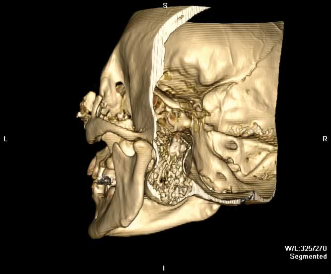
Nasoorbitoethmoid complex (NOE) fractures result from a strong blow to the midface between the orbits. NOE represents the confluence of the nasal, lacrimal, ethmoid, maxillary, and frontal bones. In addition to fractures of the paired nasal bones, fractures of the ethmoid sinus and the orbit, most commonly the lamina papyracea, are seen. NOE fractures are classified according to the degree of communition of the central bony fragment attachment site of the medial canthal tendon located in the lacrimal fossa. The medial canthal tendon is a common tendon for multiple ocular muscles. Separation of this tendon from the medial orbital wall results in lateral deviation of the globes, eyelid weakness, and watery eyes. 3D reconstructions have been shown to have a significant role in diagnosis for these fractures. Additionally, involvement of the nasofrontal ducts should be assessed, as injury can lead to mucocele formation. Severe NOE fractures can result in damage of the cribriform plate and the olfactory nerve. CSF rhinorrhea is a common clinical finding.
Scrollable Stack Images

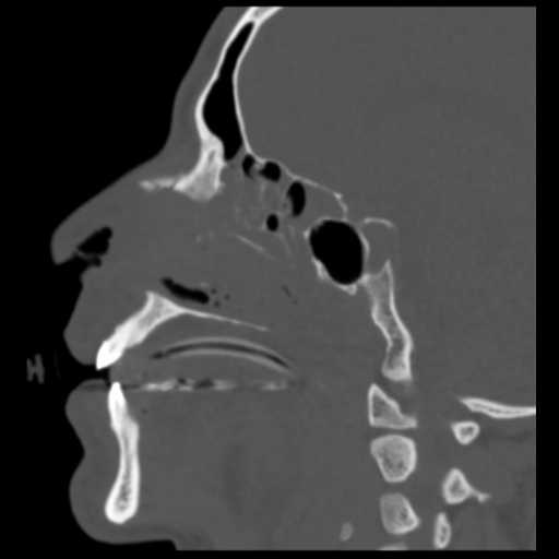
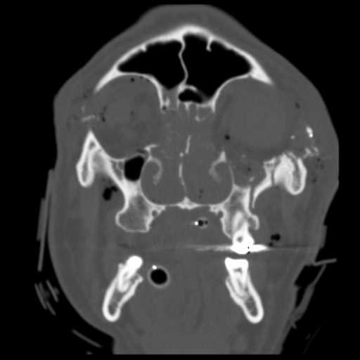
Images show complex bilateral nasoorbitoethmoid fractures. On the right, intraorbital fat herniates into the maxillary sinus through a fracture of the orbital floor. Shattering of the right lamina papyrecea can be seen. The right lateral orbital wall is shattered and the right globe is absent. Additionally, there is hemorrhagic opacification of the sphenoid sinuses. Shattering of the mid portion of the nasal septum can be seen. On the left, there are comminuted fractures of the lamina papyrecea and orbital floor.Additionally, fractures of the left lateral orbital wall, left nasolacrimal bone, all three walls of th left maxillary sinus, and shattering of the maxillary portion of the zygomatic arch are seen. The inferior rectus muscle and fat herniates into the left maxillary sinus. Vitreous hemorrhage in the left globe and a hematoma at the posteromedial aspect of the globe suggest globe rupture. Emphysema in both orbits is seen. This patient demonstrates extensive injuries in addition to nasoorbitoethmoid fractures because of a gunshot injury through the eyes.
Static 2D
 |
 |
 |
 |
| Click to enlarge | |||
Static 3D
 |
 |
 |
 |
| Click to enlarge | |||
Rotating 3D

Return to top
Axial image demonstrates shattering of the midportion of the ethmoid bone, both lamina papyrecea, and both lateral orbital walls. A minimally displaced right nasl bone fracture can be appreciated. The right globe is absent and irregularity of the left globe can be seen. There is hemorrhagic opacification of the ethmoid sinuses and bilateral intraorbital emphysema.

Return to top
A more caudal axial image demonstrates fractures of both nasal bones. Shattering of the lamina papyrecea, nasal septum, and both lateral orbital walls are seen.

Return to top
Sagittal image demonstrates nasal bone fractures and shattering of the nasal septum.

Return to top
Coronal image demonstrates shattering of the nasal septum, lamina papyrecea, and lateral orbital walls. Comminution of the left zygoma is seen. The right globe is absent and bilateral orbital emphysema is seen. There is hemorrhagic opacification of the ethmoid sinuses.

Return to top

Return to top

Return to top

Return to top
Friends
 |
Nasal Bone |
Groups
 |
Orbital Fractures |
 |
Nasal Fractures |
 |
Tripod Fractures |
 |
LeFort Fractures |
 |
Smash Fractures |
 |
Mandibular Fractures |