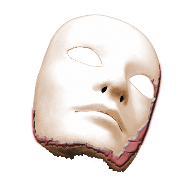
LeFort III
LeFort III fractures are transversely (coronally) oriented fractures extending superiorly from the maxilla to the zygomatic arches and posteriorly through the orbits to the pterygoid plates. This fracture pattern can be recognized by its involvement of the zygomatic arch on imaging. LeFort III fractures result in craniofacial dissociation manifested by mobility of the maxillary teeth, nose, and zygoma relate to the remainder of the skull. CSF rhinorrhea may also occur.
Scrollable Stack Images

Images show a LeFort III fracture on the left and a LeFort II fracture on the right. Minimally displaced fractures of the bilateral lateral pterygoids are seen. Fractures of the left anterior and medial maxillary walls, lateral orbital wall, orbital floor, and zygomatic arch are seen. There is significant widening of the left frontozygomatic and zygomaticotemporal sutures. LeFort II fracture on the right is seen with fracture lines running superiorly along the lateral and medial aspects of the right anterior maxillary wall. Highly comminuted fractures of the nasal bones are seen. Hemorrhagic opacificaiton of the ethmoid and maxillary sinuses are seen.
Static 2D
 |
 |
 |
 |
| Click to enlarge | |||
Static 3D
 |
 |
 |
 |
| Click to enlarge | |||
Rotating 3D

Return to top
Axial image demonstrates fractures through the lateral pterygoid plates bilaterally.

Return to top
A more cephalad axial image demonstrates fractures through the left zygomaticomaxillary and zygomaticotemporal sutures. A fracture along the left anterior maxillary wall is seen. Fractures of both medial maxillary walls with hemorrhagic opacification of the maxillary and ethmoid sinuses are seen.

Return to top
Coronal image demonstrates fractures running superiorly along the lateral and medial aspects of the left anterior maxillary wall. Nasal bone fractures can be seen.

Return to top
A more posterior coronal image demonstrates separation of the left frontozygomatic suture. Fractures through the left zygomatic arch, left maxillary sinus, and left orbital floor are seen. Fracture of the medial right maxillary wall can be appreciated.

Return to top

Return to top

Return to top

Return to top
Friends
 |
LeFort I |
 |
LeFort II |
Groups
 |
Orbital Fractures |
 |
Nasal Fractures |
 |
Tripod Fractures |
 |
LeFort Fractures |
 |
Smash Fractures |
 |
Mandibular Fractures |